Development of novel DNA marker for species discrimination of ... - Parasites & Vectors
Fasciola samples
A total of 1312 Fasciola flukes (470 F. hepatica, 609 F. gigantica, and 233 hybrid Fasciola) from 11 countries (Afghanistan, Algeria, Peru, Spain, Indonesia, Malaysia, Nigeria, Pakistan, Uganda, Japan, and Bangladesh) [8, 9, 11, 12, 14,15,16,17,18,19,20] were used in the present study. Fragment analyses of nuclear pepck and pold and the nucleotide sequencing of mitochondrial nad1 have been performed in previous studies [8, 9, 11, 12, 14,15,16,17,18,19,20]. Discrepancies between pepck and pold were observed among 7, 19, 6, 27, and 15 Fasciola isolates from Afghanistan, Algeria, Peru, Spain, and Nigeria, respectively. All available information on the Fasciola samples is summarized in Table 1.
Some of the analyses for pepck and pold were conducted in the present study. Briefly, a small portion of the vitelline glands from the posterior part of each fluke was used for DNA extraction using the High Pure PCR Template Preparation Kit (Roche, Mannheim, Germany), following the manufacturer's protocols, and stored at – 20 °C until further use. Fragments of pepck were amplified using a multiplex PCR assay with Fh-pepck-F (5′-GATTGCACCGTTAGGTTAGC-3′), Fg-pepck-F (5′-AAAGTTTCTATCCCGAACGAAG-3′), and Fcmn-pepck-R (5′-CGAAAATTATGGCATCAATGGG-3′) primers based on a previous study [7]. PCR amplicons were electrophoresed on 1.8% agarose gels for 30 min to detect fragment patterns for F. hepatica (approximately 500 bp), F. gigantica (approximately 240 bp), or hybrid (both fragments). The fragments of pold were analyzed using the PCR-RFLP assay described in a previous study [7]. The PCR products were amplified using Fasciola-pold-F1 (5′-GCTAACTTATCTGCTTACACGTGGACA-3′) and Fasciola-pold-R1 (5′-ATCGCATTCGATCAAAGCCCTCCCATG-3′) and subsequently digested with AluI enzyme (Toyobo, Osaka, Japan) at 37 °C for 3 h. The resulting products were electrophoresed on 1.8% agarose gels for 30 min to detect fragment patterns for F. hepatica (approximately 700 bp), F. gigantica (approximately 500 bp), or hybrid (both fragments).
Sequence determination of FABP type I
A primer set, FABP type I-F(5′-CACGATGGCTGACTTTGTGG-3′) and FABP type I-R(5′-AATTTTATTTGTCAGTGTTGTCGG-3′), was designed based on the mRNA sequence of FABP type I generated from F. hepatica (accession no. M95291) [13].
PCRs were performed for F. hepatica isolates from Peru, and F. gigantica isolates from Uganda in a 25 μl reaction mixture containing 2 µl template DNA, 0.2 μM of each primer, 1 U of Gflex polymerase (Takara Bio, Shiga, Japan), and the manufacturer's supplied reaction buffer. Thermal conditions included an initial denaturation step at 94 °C for 60 s, followed by 30 cycles of 98 °C for 10 s, 60 °C for 15 s, and 68 °C for 180 s. Fragments of approximately 3000 bp were amplified and purified using the NucleoSpin Gel and PCR Clean-up kit (MACHEREY–NAGEL, Düren, Germany) and then directly sequenced from both directions to obtain the preliminary sequences of FABP type I. An inner primer set, FABP type I-2F (5′-CTGGTGATGTTGAGAAGG-3′) and FABP type I-2R(5′-ACTCGTCGTCGTTTACACCCTC-3′), was generated to amplify partial FABP type I gene in F. hepatica (1951 bp) and F. gigantica (1961 bp), respectively. PCR conditions were almost the same as that described above, except for the annealing temperature, 55 °C. The nucleotide sequences of the PCR amplicons were determined precisely.
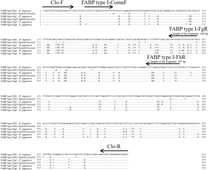
Another inner primer set, Clo-F (5′-CCATTGGTTTATAATAACTTCC-3′) and Clo-R (5′-ACTTCATTTTCTCCATCCTT-3′), which could amplify an intron of FABP type I, was designed to examine nucleotide variations between the primer regions (F. hepatica: 567 or 568 bp; F. gigantica: 566 or 567 bp) (Fig. 1). Sequence determination between Clo-F and Clo-R was performed for approximately 5% of F. hepatica and F. gigantica as well as 10 hybrid flukes selected from each country, and flukes with different nad1 haplotypes were selected as much as possible to ensure variations in the samples (Additional file 1: Table S1). PCRs were performed in a 25 μl reaction mixture containing 2 μl template DNA, 0.4 mM of each dNTP, 0.3 μM of each primer (Clo-F and Clo-R), 1 U of KOD FX Neo (Toyobo, Osaka, Japan), and the manufacturer's supplied reaction buffer. Thermal conditions included an initial denaturation step at 94 °C for 120 s, followed by 35 cycles of 98 °C for 10 s, 50 °C for 30 s, and 68 °C for 30 s. PCR products were purified using the NucleoSpin Gel and PCR Clean-up kit (Macherey-Nagel), cloned into the pUC118 Hinc II/BAP vector (Takara Bio), and sequenced. Two clones were analyzed for F. hepatica and F. gigantica, whereas four clones (two for F. hepatica genotype and two for F. gigantica genotype) were analyzed for the hybrid Fasciola fluke (Additional file 1: Table S1). The obtained sequences were aligned to construct a maximum likelihood (ML) tree using MEGA 10.0.5 software [21]. For ML tree construction, all sites were selected in the gaps/missing data treatment, and the T92 + I model was used.

Alignment of partial FABP type I gene to generate primers. Six representative genotypes of FABP type I were analyzed using Clo-F and Clo-R. A dot in the alignment indicates that the sequence was identical to that of FABP type I-Fh1 (Fasciola hepatica). The arrows indicate the position and direction of the primers. The horizontal bars represent the alignment gap. Three primers (FABP type I-ComnF, FABP type I-FhR, and FABP type I-FgR) were designed for the multiplex PCR
Multiplex PCR
A primer set for multiplex PCR was designed using the resulting sequences of Clo-F and Clo-R. FABP type I-ComnF (5-′GCGGTTCTGAGTGTGTGTTT-3′) is a common primer for F. hepatica and F. gigantica, whereas FABP type I-FhR (5′-TGACGAACAGCTTACCTTCGAG-3′) and FABP type I-FgR (5′-CAATACTCCTCACCACCCAG-3′) are specific to F. hepatica (length of the amplicon: 287 bp) and F. gigantica (189 bp), respectively (Fig. 1). PCR amplification was performed in 10 μl reaction mixtures containing 0.5 µl template DNA, 0.1 µM of each dNTP, 0.2 µM of each primer, 0.01 U of Go Taq DNA Polymerase (Promega, Madison, WI, USA), and the manufacturer's supplied reaction buffer. The PCR conditions included an initial denaturation step at 95 °C for 120 s, followed by 35 cycles of 95 °C for 30 s, 55 °C for 30 s, and 72 °C for 60 s, and a final extension step at 72 °C for 5 min. PCR amplicons were electrophoresed on 1.8% agarose gels and visualized using ethidium bromide staining. Multiplex PCR was then applied to all 1312 flukes (Table 1).
Comments
Post a Comment